
- Resource
- BR1DGE
- Early Detection
- Article
IDS 2026 Symposium Summary
In this panel Prof. Chantal Mathieu facilitated the discussion with Prof. Ezio Bonifacio, Prof. Thomas Kay, and Prof. John Wentworth. The speakers discussed biomarker-driven type 1 diabetes care, focusing on screening, diagnosis, and monitoring using autoantibodies, C-peptide, and glucose metrics. The panel highlighted the importance of early detection and risk stratification, reviewed immune-targeted therapies and emerging technologies, and emphasized key decision points supporting personalized, biomarker-guided T1D management.
This article is intended to be a summary only. Additional topics were covered in the live symposium in line with the applicable regulations but are not included on the BR1DGE platform.

Meeting Objectives
- Examine the evolving biomarker landscape in T1D, including autoantibodies, genetic risk scores, cellular immunity markers, and emerging multi-omic signatures that define disease progression
- Explore advances in immunomodulatory approaches and how understanding of immune pathways is shaping knowledge of potential disease modification in T1D
- Discuss clinical translation of biomarker science through early detection programs and their role in optimizing the care of individuals with early-stage T1D
Symposium Faculty




The Immune Architecture of T1D and Potential Therapeutic Targets

“I would say that most of these treatments need to directly or indirectly affect T cells because we have recognised that they sit at the central part of the pathogenesis of the disease.”
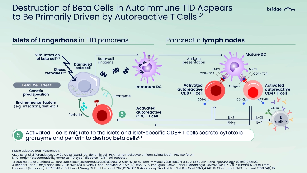
- Type 1 diabetes has a complex multifactorial etiology driven by genetic predisposition and environmental triggers, leading to autoimmunity and autoreactive T cell attack on pancreatic beta cells in the islets of Langerhans1-3
- Dendritic cells play a key role by capturing beta-cell antigens in islets, migrating to pancreatic lymph nodes, activating CD4+ and CD8+ T cells and contributing B cell activation and antigen presentation4-9
- B cells capture self-antigens to boost T cell activation, and CD4⁺ T cells enhance antibody production and support CD8⁺ T cell responses. Meanwhile CD8⁺ T cells drive beta-cell destruction by releasing IFN-γ and producing perforin and granzyme as the primary mechanism of beta-cell death4,6,8,10-11
- In T1D, a critical imbalance between immune activation and immune regulation, including T regulatory cell pathways and T cell exhaustion, drives disease progression4,6,12-16
- Disease-modifying therapies targets multiple disease stages through strategies that modulate autoreactive T cells, regulatory T cells, B cells, inflammatory cytokines, and beta cell protection17-22
- Consistent with therapeutic strategies targeting autoreactive T cells, a Phase 2 ongoing trial in people with Stage 3 T1D is evaluating multiple doses of an immunotherapeutic agent over 12 months with a blinded 12-month extension phase, with primary phase completion expected in approximately 12 months.23 Primary outcome is the change from baseline to Week 52 in mean 2-hour MMTT stimulated C-peptide concentration, and secondary outcomes include efficacy through Weeks 52 and 104, as well as safety, tolerability, pharmacokinetics of the immunotherapeutic agent, and patient-/caregiver-reported clinical outcomes23,24
- Achieving robust and durable outcomes in T1D requires matching therapies to targets, using immune biomarkers to predict therapeutic effect, considering extended interventions to overcome immune escape mechanisms, and exploring combination therapies to address the heterogeneous nature of T1D18,19,25,26
Immune Biomarkers: Enabling Detection and Providing Insights into Disease Pathophysiology

“If you have a C-peptide, your diabetes is better... keeping and having C-peptide is a good thing... the more you preserve it, the lower the HbA1c remains.”
0.png/jcr:content/image%20(2).png)
- Type 1 Diabetes is uniquely advantaged among autoimmune conditions in having well-established biomarkers — four islet autoantibodies (IAA, GAD, IA-2, ZnT8), with the consistent presence of two or more in children conferring a lifetime risk of T1D approaching 100%1-2
- Early detection and monitoring of T1D through islet autoantibody screening is associated with improved HbA1c, fasting glucose, and C-peptide levels, reduced DKA risk, clinical trial enrollment opportunities, and more structured insulin therapy initiation and diabetes education and counseling3-12
- Key antibody characteristics associated with faster progression include higher numbers of detected autoantibodies (≥2), higher titer levels, early appearance, and specific antibody type — with IA-2 autoantibody positivity identified as one of the strongest predictors of rapid progression and closest to a fast-progressor endotype1, 13-23
- The number and type of autoantibodies significantly influence T1D risk, with single transient antibodies carrying the lower risk and the early presence of multiple IA2-containing antibodies is associated with higher risk and increased disease progression16,23
- C-peptide is the preferred biomarker for beta-cell function due to its constant clearance rate, longer half-life (20–30 minutes), and negligible hepatic extraction, making it more reliable than insulin for plasma measurement24-30
- Clinical data from INNODIA, Scottish registries, a cross-sectional study, and TOMI-T1D pooled analyses confirm that preserved C-peptide levels correlate with better glycemic control (lower HbA1c, improved CGM values) and support its use as a sensitive and clinically meaningful endpoint, often enabling more efficient and statistically robust trial designs31-35
Investigating How Biomarkers Can Inform Risk Prediction in T1D

“The OGTT is pretty good, and you know of course it will predict who is going to go to Stage 3 diabetes because we are measuring the outcome that we use to diagnose and define Stage 3… But it does have quite major limitations, particularly in the setting of children… I think particularly in paediatrics we are going to need to start applying this but also other risk scores to actually find the kids who are going to benefit most from immune-targeted interventions.”
.png/jcr:content/image%20(3).png)
- Risk stratification in islet autoimmunity is improved by combining OGTT results with IA2 antibody status, age, and BMI, enabling better prediction of progression to Stage 3 diabetes1
- A range of T1D risk prediction scores are currently available; some of these scores include the M120 score, the Progression Likelihood Score (PLS), and the Combined Risk Score (CRS), developed to improve prediction of progression to Stage 3 T1D beyond what the OGTT alone provides1–7
- The M120 score, developed using TrialNet Natural History data and validated across multiple cohorts (TrialNet, TEDDY, DPT-1, FR1DA), incorporates 120-minute post-OGTT glucose, C-peptide, HbA1c, age-adjusted BMI, and IA-2 autoantibody status into a single time-point measure1
- Use of the M120 threshold significantly improved identification of high-risk status for progression to Stage 3 clinical T1D in children with multiple autoantibodies, particularly among Stage 1 individuals considered for pediatric immune interventions1
- The M120 score was also used as a surrogate marker of beta cell function in the TN10 trial, where it detected a significant treatment effect at both months three and six versus baseline1
- The Progression Likelihood Score (PLS), derived from the Fr1da study, integrates metabolic variables (90-min glucose and HbA1c) and immunologic variables (IA-2A status and titer [low, intermediate and high]) to predict the risk of T1D progression. When applied to the Fr1da cohort, only a minority of children in Stage 1 had a high progression score, but those identified were at high risk of progression to Stage 3 within two years2
- The Combined Risk Score (CRS), derived from the TEDDY data, uses Genetic Risk Score 2 (GRS2), family history of T1D, and antibody count, but its clinical use is limited by the requirement for GRS2 genotyping; incorporating metabolic inputs and IA-2 status could further improve its performance3
MAT-GLB-2603716 – 1.0 – 07/2026
Early Detection and Screening
1. Primavera M, et al. Front Endocrinol (Lausanne). 2020;11:248.
2.Verduci E, et al. Front Nutr. 2020;7:612377.
3. American Diabetes Association. Diabetes Care. 2025;48(Suppl.1):S27-49.
4. Houeiss P, Luce S, Boitard C. Front Endocrinol (Lausanne). 2022;13:933965
5. Li Y, et al. Front Immunol. 2021;12:690783.
6. Burrack AL, et al. Front Endocrinol (Lausanne). 2017;8:343.
7. Boldison J, Wong FS. Front Immunol. 2021;12:746187.
8. Addissouky TA, et al. Bull Natl Res Cent. 2024;48:42.
9. Choi H, et al. BMC Immunol. 2023;24(1):15
10. Richardson SJ, et al. Diabetologia. 2016;59(11):2448-58.
11. den Hollander NHM, Roep BO. Front Med (Lausanne). 2022;9:932086
12. Garg G, et al. J Immunol. 2012;188(9):4644-53.
13. Quattrin T, Mastrandrea LD, Walker LSK. Lancet. 2023;401(10394):2149-62.
14. Lindley S, et al. Diabetes. 2005;54(1):92-9.
15. Schneider A, et al. J Immunol. 2008;181(10):7350-5.
16. Kwong C-T, et al. Immun Cell Biol. 2021;99:486-95.
17. Besser REJ, et al. Pediatr Diabetes. 2022;23(8):1175-8
18. Herold KC, et al. Nat Rev Immunol. 2024;24(6):435-51
19. Zarei M, et al. Diabetes Epidemiol Manage. 2025;17:100246.
20. Jacobsen LM, et al. Curr Diab Rep. 2018;18(10):90
21. Li Y, et al. Front Immunol. 2021;12:690783
22. Weiskorn J, et al. Horm Res Paediatr. 2025;98(5):558-65
23. ClinicalTrials.gov. NCT06111586. Accessed March 2026.
24. INNODIA. FABULINUS Trial Information. Available at: https://www.innodia.org/fabulinus-trial. Accessed March 2026
25. Ziegler AG, et al. Lancet. 2025;406(10511):1520-34
26. Brenu EW, et al. Front Endocrinol. 2023;14:1117076.
Immune Biomarkers: Enabling Detection and Providing Insights into Disease Pathophysiology
1. Insel RA, et al. Diabetes Care. 2015;38(10):1964-74.
2. ADA 2026 Standard of Care Guidelines. Diabetes Care. 2026;49:S1-S371
3. Hummel S, et al. Diabetologia. 2023;66:1633-42.
4. Latres E, et al. Diabetes 2024;73:823-33.
5. Phillip M, et al. Diabetologia. 2024;67(9):1731-59.
6. Ziegler AG, et al. JAMA. 2020;323:339-51.
7. Steck AK, et al. Diabetes Care. 2022;45(2):365-71.
8. Leichter SB, et al. J Clin Endocrinol Metab. 2025;110:2371-82
9. Bendor-Samuel OW, et al. Arch Dis Child. 2023;108(1):26-30.
10. Kick K, et al. Contemp Clin Trials Commun. 2018:11:170-73
11. Oron T, et al. Pediatr Diabetes. 2024:4238394.
12. Bonifacio EB, et al. Diabetes Obes Metab. 2025;27(Suppl.6):28-39
13. Ziegler AG, et al. JAMA. 2013;309:2473-79.
14. Orban T, et al. Diabetes Care. 2009;32:2269-74.
15. Bonifacio E. Diabetes Care. 2015;38:989-96.
16. Anand V, et al. Diabetes Care. 2021;44(10):2269-76.
17. Frohnert B, et al. Diabetes Care. 2023;46(10):1753-61.
18. Felton JL, et al. Commun Med (Lond). 2024;4(1):66.
19. So M, et al. Endocr Rev. 2021;42(5):584-604
20. Jacobsen LM, et al. Diabetologia. 2020;63(3):588-96.
21. Pöllänen PM, et al. Diabetologia. 2017;60(7):1284-93.
22. Suomi T, et al. EBioMedicine. 2023;92:104625.
23. Ghalwash M, et al. Diabetes Care. 2024;47(8):1424-31
24. Mameli C, et al. Pharmacol Res. 2023;193:106792.
25. Leighton E, et al. Diabetes Ther. 2017;8(3):475-87.
26. Palmer JP, et al. Diabetes 2004;53(1):250-64.
27. Galderisi A, et al. Diabetologia. 2023;66(12):2189-99.
28. Flier JS, Kahn CR. Mol Metab. 2021;52:101194.
29. Piccinini F, Bergman RN. Diabetes Care. 2020;43(9):2296-302.
30. Matteucci E, et al. Drug Des Devel Ther. 2015;9:3109-18.
31. Marcovecchio ML, et al. Diabetologia. 2024;67(6):995-1008
32. Jeyam A, et al. Diabetes Care. 2021;44(2):390-8.
33. Stimson RH, et al. Diabetologia. 2026;69(1):59-68.
34. Taylor PN, et al. Lancet Diabetes Endocrinol. 2023;11(12):915-25.
35. Latres E, et al. Diabetes. 2024;73(6):823-33
Investigating How Biomarkers Can Inform Risk Prediction in T1D
1. Le MV, et al. Diabetes Care. 2025;48(8):1352-5.
2. Weiss A, et al. Diabetologia. 2022;65(12):2121-31
3. Ferrat LA, et al. Nat Med. 2020;26(8):1247-55.
4. Sosenko JM, et al. Diabetes Care. 2013;36(9):2615–20.
5. Simmons KM, et al. J Clin Endocrinol. 2020;105(11):e4094–e4101.
6. Jacobsen LM, et al. J Clin EndocrinolMetab. 2022;107(10):2784-92.
7. Joglekar MV, et al. 2025;31(8):2622-31.